Last Updated on October 4, 2025 by Aaron Barriga
Our eyes are integral to our body, allowing us to view the world around us. Imagine being able to touch, smell, hear, and feel everything but not see, like a car with dead headlights driving down a highway. The dark isolation of vision loss can be quite horrifying!
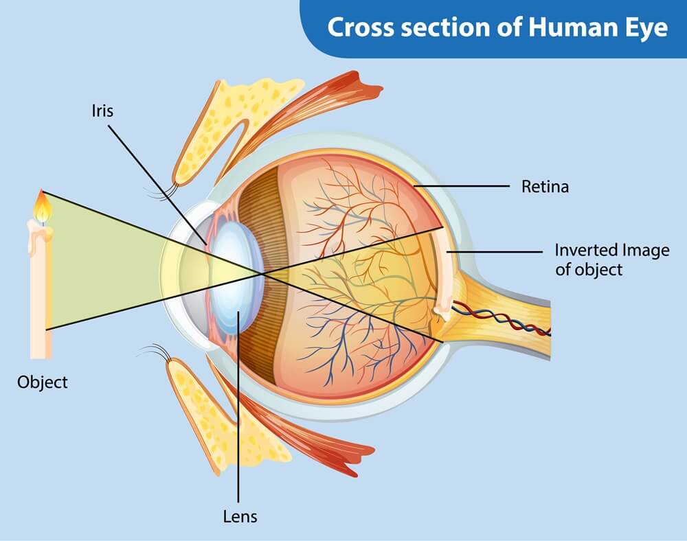
This fantastic process happens when the optic nerve creates an image through the cornea and displays it on the retina (a light-sensitive layer of tissue that acts like a screen). Retinal detachment, or RD, is a medical condition where the retina peels off from the surrounding tissue.
What are the Types of Retinal Detachment?
 There are three types of retinal detachment:
- Rhegmatogenous: The most common type, where the retina detaches from the Retina Pigment Epithelium (RPE) due to fluid getting under it after a retinal tear.
- Tractional: A rare but serious type, where the cracked retina rips from the RPE due to the contracting scar tissue on its surface.
- Exudative: This type occurs without holes, tears, or breaks in the retina, and is caused by fluid leaking into the area under it (typically due to inflammation, injury, or vascular abnormalities).
What Causes Retinal Detachment?
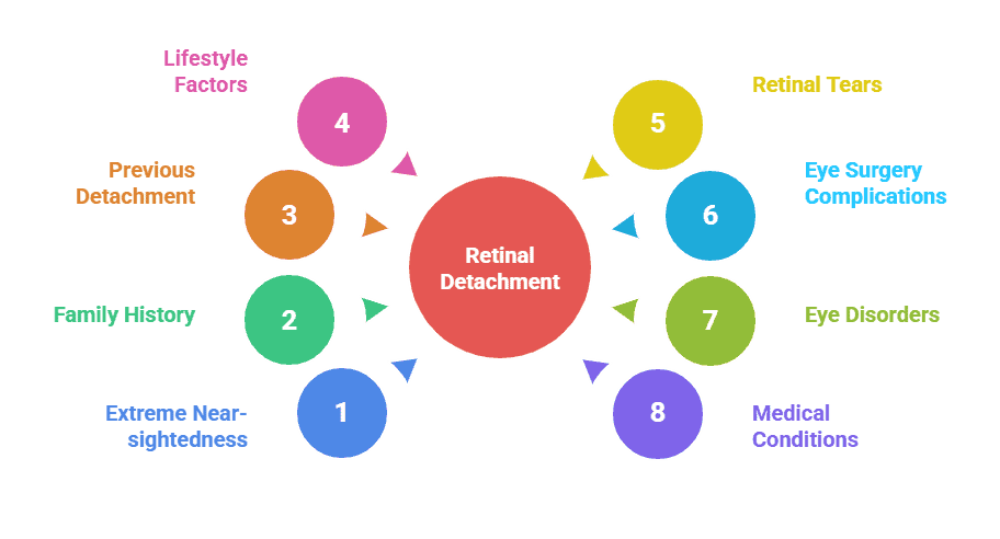 Here are some of the common risk factors for Retinal Detachment:
- Extreme near-sightedness
- Family history of Retinal Detachment
- Previous retinal detachment in one eye
- Stress and lifestyle factors like smoking
- Retinal tears caused by injury to the eyes or head
- Complications after an eye surgery, e.g., cataract surgery
- Tumors, degenerative myopia, lattice degeneration, glaucoma, and other eye disorders
- Other diseases and medical conditions like diabetes, AIDS, sickle cell disease, etc.
Who’s at Risk of Retinal Detachment?
RD caused due to injury or underlying medical conditions can happen at practically any age, but is more likely to occur after the age of 40. Almost half of all the people who have retinal tears are likely to end up with a detached retina, which puts sportspeople and athletes at high risk. Men are more prone to it than women, and research shows that Caucasians are at higher risk than African Americans.
Also Read: 11 Common Eye Problems in Old Age
Symptoms and Signs of Retinal Detachment
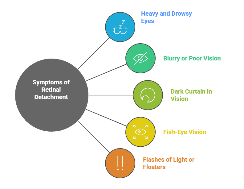 There is no pain associated with retinal tears, but at the initial stage, there are certain signs that can help with the detection of Retinal Detachment. These include:
- Eyes are becoming heavy and drowsy
- Blurry or poor vision, a thin layer or ‘shadow’ hindering sight
- A dark ‘curtain’ approaching the center of your vision from the side (peripheral vision).
- Fish-eye vision for linear objects like roads, trees, buildings, etc.
- Sudden flashes of light or ‘floaters’ in your vision
Treatment and Cure of Retinal Detachment
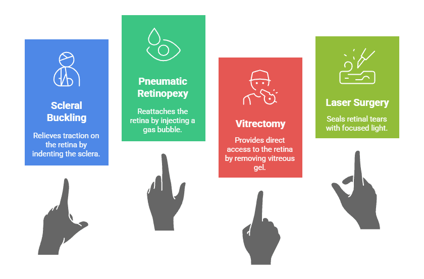 RD is curable if the breaks in the retina can be closed or sealed again. An ophthalmologist can use fundus photography or ophthalmoscopy to diagnose it and conduct various surgical procedures for treatment. These include:
- Scleral Buckling: Of the four types of retinal detachment surgery, this is the most popular one. Small bands of silicone or plastic are attached to the outside portion of the eye – the sclera. As a result which the band compresses the eye inward, which minimizes the traction of the retina, which eventually allows it to reattach itself to the inner wall of the eye. The scleral buckle thus created attaches to the posterior surface of the eye but is invisible long after this surgery is completed. In some cases, the scleral buckling surgery may be performed in combination with other surgical procedures so as to fuse the retina with the retinal pigment epithelium, the underlying supportive tissue.
- Pneumatic Retinopexy: In this surgical procedure, a small gas bubble is injected into the vitreous body, the gel-like substance present between the lens and the retina. Because of this bubble, the vitreous rises and presses against the retina, which effectively closes the tear. After this, a freezing probe of a laser can be used to seal tears on the retina. This use of a freezing probe is termed cryopexy, and the usage of a laser beam is known as photocoagulation. However, the rate of success of this retinal detachment surgery is lower than the Scleral Buckling procedure.
- Vitrectomy: This retinal detachment surgical procedure is becoming increasingly popular. In this treatment, the clear jelly-like fluid from the vitreous body, which is the posterior chamber of the eye. Silicone oil is then used to replace this fluid to push the detached part of the retina into the correct position.
- Laser Surgery: In this procedure, a laser beam is used to burn the area around the tear to create scarring on the retina’s underlying tissue.
In addition to the above procedures, Cryotherapy or laser photocoagulation may be used to prevent the detachment from spreading.
In some cases, the surgical reattachment of the retina may not be successful because of certain factors such as the extent, cause, and area of detachment. Some complications that may develop during or after the surgery are as follows:
- Increased tears on the retina
- Bleeding in the eye
- High pressure or swelling in the eye
- Double vision
- A rare possibility of infection in the eye
- Bruising around the eye
- Allergic reaction to the medication
- Cloudiness in the lens of the eye
Can RD Be Prevented?
To prevent RD, be proactive and tackle the initial stage symptoms. Schedule a regular eye checkup every year and consult your eye doctor if you suspect a retinal tear or rip. Also, wear protective goggles for sports, driving, working with chemicals, machines, or tools, etc.
It Can Happen to Anyone!
There are some famous personalities who suffered from retinal detachment, including the 26th American President Theodore Roosevelt, renowned basketball player Amar’e Stoudemire, noted publisher, journalist, and politician Joseph Pulitzer, soccer star Pele, and various others.
Vision loss caused by RD can be prevented or cured with early treatment, if the retinal tear is corrected before detachment occurs. RD is considered a medical emergency that can severely affect your eyesight or even lead to blindness if left untreated, so if you have the symptoms, get in touch with an experienced ophthalmologist or eye surgeon at the InSight Vision Center!
Don’t wait until it’s too late—if you’re experiencing symptoms of retinal detachment, book an appointment with our expert eye surgeons at InSight Vision Center today!

Dr. Azhar I. Salahuddin is an ophthalmologist and is fellowship-trained in cornea, external diseases, and refractive surgery. Dr. Salahuddin has been performing cataract surgery for over 19 years and specializes ocular reconstruction, corneal transplantation surgery as well as vision correction through a variety of intraocular lenses. Dr. Salahuddin is board-certified by the American Board of Ophthalmology and was trained at Boston University.